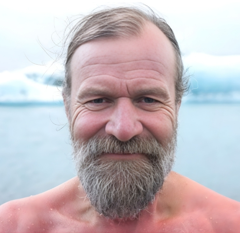
“The Iceman” Wim Hof
Wim Hof holds 26 Guinness World Records ranging from cold endurance challenges such as climbing Mount Kilimanjaro within two days to running a full marathon (42 kilometres (26 miles)) above the Arctic Circle at temperatures hovering around −20 °C (−4 °F). Mr. Hof would accomplish both of these feats wearing nothing but a pair of shorts. In 2011, Wim would also complete a 26 mile marathon in the Namib Desert in Africa without ingesting any water.
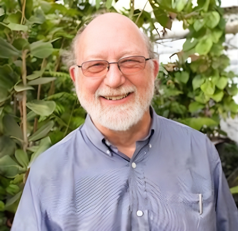
Dr. Dennis Mckenna
Dr. Dennis McKenna is an ethnopharmacologist, author and founder of Symbio Life Sciences. He has dedicated his career to the study of hallucinogens and psychedelics and is a founding board member of the Heffter Research Institute, a non-profit organisation dedicated to investigating therapeutic uses of psychedelic medicines.
Dr. Jon Dean
Dr. Jon Dean is a postdoctoral scientist in the Brain Mechanisms of Pain and Health Laboratory at UC San Diego’s Department of Anesthesiology. After obtaining his B.A. in Chemistry from Youngstown State University, Dr. Dean completed his Ph.D. in Molecular and Integrative Physiology from the University of Michigan.
Dr. Steven Barker
Dr. Steven Barker is a pioneering pharmacologist and chemist who has focused his career on psychedelic research, particularly N,N-dimethyltryptamine (DMT), beginning his investigations in 1976. He earned his Ph.D. from the University of Alabama at Birmingham in 1978 with a dissertation titled “N,N-Dimethyltryptamine – An Endogenous Hallucinogen” and went on to become Professor Emeritus at Louisiana State University’s School of Veterinary Medicine, where he directed the Analytical Systems Laboratory and served as State Chemist for the Louisiana State Racing Commission while holding the Everett D. Besch Distinguished Professor award from 2000 to 2006.
Anton Bilton
Anton Bilton is an international property entrepreneur, philanthropist, and a consciousness researcher. In 2015, Anton launched the Tyringham Initiative (TI) as a world-class think-tank for the evolution, expansion and deeper understanding of the human experience and potential.
Dr. Ede Frecksa
Dr. Ede Frecska is the Chairman of Psychiatry at the Faculty of Medicine of the University of Debrecen. He received his medical degree in 1977 from the Semmelweis University in Hungary. He then earned qualifications as certified psychologist from the Department of Psychology at Lorand Eotvos University in Budapest. Dr. Frecska completed his residency training in Psychiatry both in Hungary (1986).
Dr. Mauro Zappaterra
Dr. Zappaterra graduated from Harvard Medical School with an MD and PhD and now specializes in Physical Medicine and Rehabilitation with a particular focus on regenerative medicine, neuro-rehabilitation, neuroplasticity, musculoskeletal medicine, and pain. He completed his PhD doing work with neuronal stem cells and the effects of the cerebrospinal fluid (CSF) in brain development and in the adult.
Project Founder
John Chavez - I’m just a guy that believes that distinct changes in perception and “supernormal” abilities can be converged into a demystified conversation. Based on my experience, I do believe the world can change quickly and profoundly once the subject has been developed to its fullest potential. In 2026, the connectivity of inspired minds across the world will assist in creating a new conversation about what we think we know about this world and our places in it.